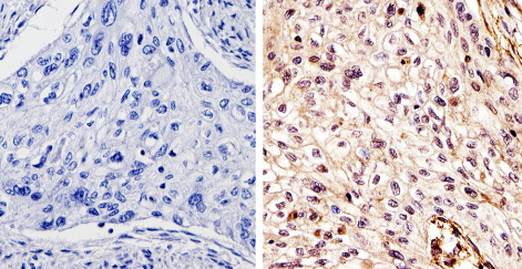
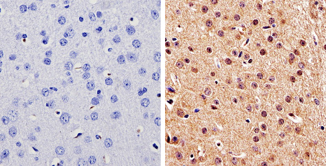
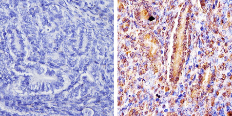
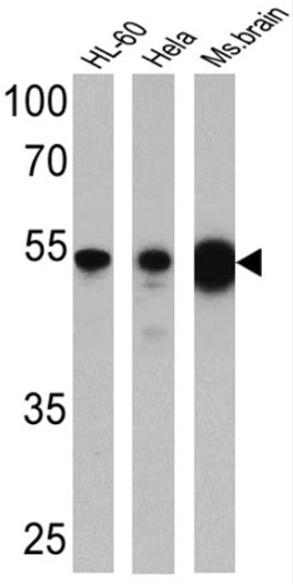
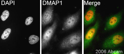
Anti-DMAP1 antibody - ChIP Grade
| Name | Anti-DMAP1 antibody - ChIP Grade |
|---|---|
| Supplier | Abcam |
| Catalog | ab2848 |
| Prices | $380.00 |
| Sizes | 100 µl |
| Host | Rabbit |
| Clonality | Polyclonal |
| Isotype | IgG |
| Applications | ChIP IHC-P WB ICC/IF ICC/IF |
| Species Reactivities | Mouse, Human, Xenopus |
| Antigen | Synthetic peptide corresponding to Mouse DMAP1 aa 435-450 |
| Description | Rabbit Polyclonal |
| Gene | DMAP1 |
| Conjugate | Unconjugated |
| Supplier Page | Shop |
Product images
Product References
Knockout of mouse Cyp3a gene enhances synthesis of cholesterol and bile acid in - Knockout of mouse Cyp3a gene enhances synthesis of cholesterol and bile acid in
Hashimoto M, Kobayashi K, Watanabe M, Kazuki Y, Takehara S, Inaba A, Nitta S, Senda N, Oshimura M, Chiba K. J Lipid Res. 2013 Aug;54(8):2060-8.
Distinct roles of DMAP1 in mouse development. - Distinct roles of DMAP1 in mouse development.
Mohan KN, Ding F, Chaillet JR. Mol Cell Biol. 2011 May;31(9):1861-9.
DNA methyltransferase 1-associated protein (DMAP1) is a co-repressor that - DNA methyltransferase 1-associated protein (DMAP1) is a co-repressor that
Lee GE, Kim JH, Taylor M, Muller MT. J Biol Chem. 2010 Nov 26;285(48):37630-40.
Dmap1 plays an essential role in the maintenance of genome integrity through the - Dmap1 plays an essential role in the maintenance of genome integrity through the
Negishi M, Chiba T, Saraya A, Miyagi S, Iwama A. Genes Cells. 2009 Nov;14(11):1347-57.
Bmi1 cooperates with Dnmt1-associated protein 1 in gene silencing. - Bmi1 cooperates with Dnmt1-associated protein 1 in gene silencing.
Negishi M, Saraya A, Miyagi S, Nagao K, Inagaki Y, Nishikawa M, Tajima S, Koseki H, Tsuda H, Takasaki Y, Nakauchi H, Iwama A. Biochem Biophys Res Commun. 2007 Feb 23;353(4):992-8. Epub 2006 Dec 29.
