Anti-DMT1 antibody (ab123085) at 1/100 dilution + Mouse cerebellum tissue lysates at 35 µg

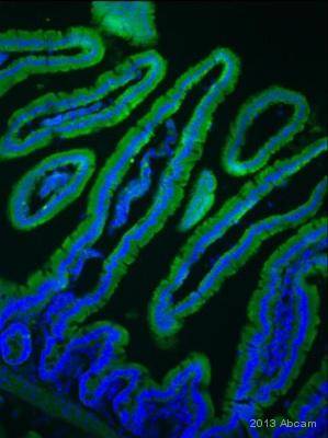
IHC-FoFr image of DMT1 staining on mouse intestine tissue using ab123085 (1:100). Adult mouse was perfused with 4% PFA in 0.1M PBS. The sections were cut using a cryostat and permeabilized using 0.1% TritonX in 0.1% PBS. The sections were then blocked using donkey serum for 1 hour at 24°C. ab123085 (1:100) was incubated with the sections for 24 hours at 4°C. The secondary antibody used was donkey polyclonal to mouse IgG conjugated to alexa fluor 488 (1:1000). See Abreview

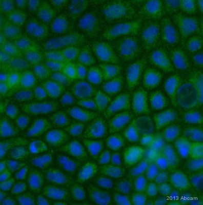
ICC/IF image of DMT1 staining on Caco2 cells using ab123085 (1:100). The cells were fixed using paraformaldehyde and permeabilized using 0.1%TritonX in 0.1 %PBS. The cells were blocked using 10% donkey serum for 1 hour at 24°C. ab123085 (1:100) was incubated with the cells for 4 hours at 25°C. The secondary antibody used was donkey polyclonal to Rabbit IgG conjugated to Alexa Fluor 488.See Abreview


