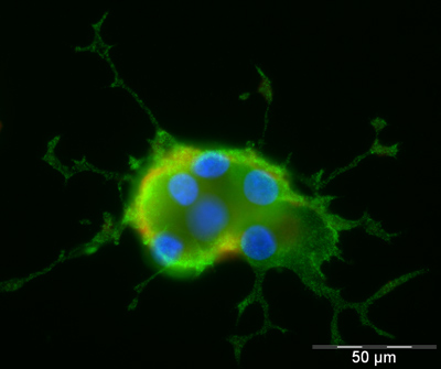
ICC/IF image of ab28941 stained PC12 cells. The cells were 4% formaldehyde fixed (10 min) and then incubated in 1%BSA / 10% normal goat serum / 0.3M glycine in 0.1% PBS-Tween for 1h to permeabilise the cells and block non-specific protein-protein interactions. The cells were then incubated with the antibody (ab28941, 1µg/ml) overnight at +4°C. The secondary antibody (green) was Alexa Fluor® 488 goat anti-rabbit IgG (H+L) used at a 1/1000 dilution for 1h. Alexa Fluor® 594 WGA was used to label plasma membranes (red) at a 1/200 dilution for 1h. DAPI was used to stain the cell nuclei (blue).

Doublecortin antibody - Neuronal Marker (ab28941; 5ug/ml) cytosolic and axonal staining in dorsal root ganglion explants, dissected from 16 day-old rat embryos and cultured for 6 hours or 4 days in vitro with Neurobasal Medium containing 10% fetal calf serum and B27 supplement. Immunocytochemistry: All steps were performed in PBS. Cells or explants were fixed in 4% PFA for 15min, permeabilised with 0.1% TX100 for 10min and blocked with 5% BSA, 0.1% TX100 for 45min. ab28941 was incubated at 12h in 5% BSA, 0.1% TX100 at 4°C. Preincubation of ab28941 with immunising peptide ab19803 blocked immunostaining. To-pro-3 was used as a nuclear counterstain. Treated cultures were mounted on glass coverslips with Mowiol.

All lanes : Anti-Doublecortin antibody - Neuronal Marker (ab28941) at 1 µg/mlLane 1 : Mouse brain lysateLane 2 : Mouse brain lysate with Rat Doublecortin peptide (ab19803) at 1 µg/mlLysates/proteins at 20 µg per lane.SecondaryAlexa fluor goat polyclonal to IgG (700) at 1/10000 dilutionPerformed under reducing conditions.


