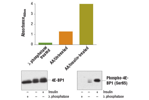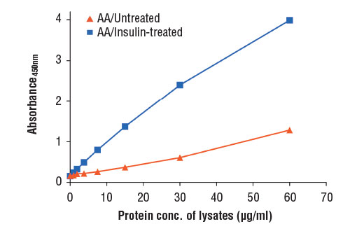
Figure 1: Treatment of HEK-293T cells with amino acids and insulin stimulates phosphorylation of 4E-BP1 at Ser65, as detected by PathScan ® Phospho-4E-BP1 (Ser65) Sandwich ELISA kit #13777, but does not affect the level of total 4E-BP1 protein detected by western analysis. HEK-293T cells (70-80% confluent) were starved overnight and deprived of amino acids for 1 hr. The amino acids were replenished for 1 hr. Cells were either untreated (-) or stimulated with 100 nM insulin for 30 min at 37 o C (+). Treatment of control cell lysates with λ phosphatase (4000 U/ml, 60 min, 37 o C) abolishes the basal phosphorylation of 4E-BP1 as shown by both sandwich ELISA and western blot analysis. The absorbance readings at 450 nm are shown in the top figure, while the corresponding western blots, using 4E-BP1 (53H11) Rabbit mAb #9644 (left panel) or Phospho-4E-BP1 (Ser65) (174A9) Rabbit mAb #9456 (right panel), are shown in the bottom figure.

