 Immunohistochemistry
Rabbit Anti-Phospho MCM2 (S40/S41) Antibody, Affinity Purified:
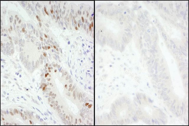
Cat. No. PLA0141: Detection of Human Phospho MCM2 (S40/S41) by Immunohistochemistry. Samples: FFPE serial sections of Human colon carcinoma. Mock phosphatase treated section (left) and calf intestinal phosphatase-treated section (right). Antibody: Affinity purified Rabbit Anti-Phospho MCM2 (S40/S41) (
Cat. No. PLA0141) used at a dilution of 1:200 (1 μg/mL). Detection: DAB
 Immunofluorescence
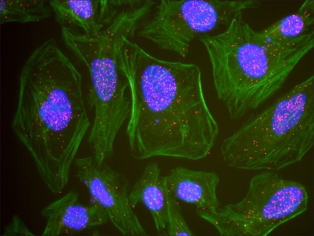
: This antibody is qualified for the Proximity Ligation Assay (PLA). The above image shows representative results for PLA using three color fluorescence, including DAPI stained nuclei (blue), phalloidin stained cytoplasmic F-actin (green), and PLA positive signal (red).
 Immunoblotting
Rabbit Anti-Phospho MCM2 (S40/S41) Antibody, Affinity Purified:
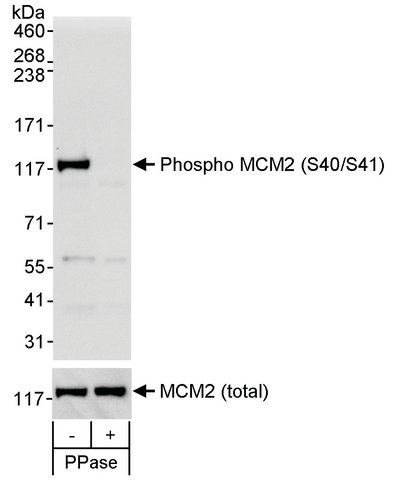
Cat. No. PLA0141: Detection of Human Phospho MCM2 (S40/S41) by Western Blot. Samples: Whole cell lysate (50 μg) from asynchronous 293T cells that was mock treated (-) or treated (+) with phosphatases (PPase). Antibody: Affinity purified Rabbit Anti-phospho MCM2 (S40/S41) Antibody (
Cat. No. PLA0141) used at 0.1 μg/mL. To examine total MCM2, the blot was stripped and then blotted with Rabbit Anti-MCM2 Antibody (
Cat. No. PLA0060) at 0.1 μg/mL. Detection: Chemiluminescence with exposure times of 30 seconds (upper and lower panels).
![<B>Flow Cytometry</B><BR/>Rabbit Anti-Phospho MCM2 (S40/S41) Antibody, Affinity Purified: <B>Cat. No. PLA0141</B>: Flow cytometric analysis of phospho-MCM2 (pS40/41). Jurkat cells were fixed in 1.5% PFA, and permeabilized in 90% MeOH. 1 × 10<SUP>6</SUP> cells were stained with 0.1 μg/mL Anti-phospho-MCM2 (pS40/41) [<B>PLA0141</B>] in a 150 mcl volume. DNA content was simultaneously analyzed via PI stain.](http://www.bioprodhub.com/system/product_images/ak_products/7/sub_1/3567_pla0141-facs-1-large.jpg) Flow Cytometry
Flow CytometryRabbit Anti-Phospho MCM2 (S40/S41) Antibody, Affinity Purified:
Cat. No. PLA0141: Flow cytometric analysis of phospho-MCM2 (pS40/41). Jurkat cells were fixed in 1.5% PFA, and permeabilized in 90% MeOH. 1 × 10
6 cells were stained with 0.1 μg/mL Anti-phospho-MCM2 (pS40/41) [
PLA0141] in a 150 mcl volume. DNA content was simultaneously analyzed via PI stain.


![<B>Flow Cytometry</B><BR/>Rabbit Anti-Phospho MCM2 (S40/S41) Antibody, Affinity Purified: <B>Cat. No. PLA0141</B>: Flow cytometric analysis of phospho-MCM2 (pS40/41). Jurkat cells were fixed in 1.5% PFA, and permeabilized in 90% MeOH. 1 × 10<SUP>6</SUP> cells were stained with 0.1 μg/mL Anti-phospho-MCM2 (pS40/41) [<B>PLA0141</B>] in a 150 mcl volume. DNA content was simultaneously analyzed via PI stain.](http://www.bioprodhub.com/system/product_images/ak_products/7/sub_1/3567_pla0141-facs-1-large.jpg)