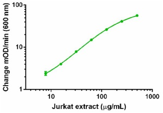
A dilution series of Jurkat cell extract in Incubation Buffer in the working range of the assay.

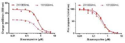
Lysates corresponding to 2x10⁶ cells/mL or 1x10⁶ cells/mL were prepared by direct in-well lysis (without media removal) from Jurkat cells treated with variable doses of staurosporine for 6 hours in a 96-well plate. 100 µL of the lysates were analyzed. Background-subtracted signals are shown in left panel. Relative pro caspase 3 concentrations interpolated from standard curve of Jurkat cell extract dilutions and expressed in percent of vehicle-treated control are shown in right panel. IC50 were determined from both absolute signals (0.60 µM and 0.42 µM using, respectively, 2x10⁶ cells/mL and 1x10⁶ cells/mL) and relative signals (0.42 µM and 0.35 µM using, respectively, 2x10⁶ cells/mL and 1x10⁶ cells/mL). Note close match of IC50 value determined from absolute and relative signals.

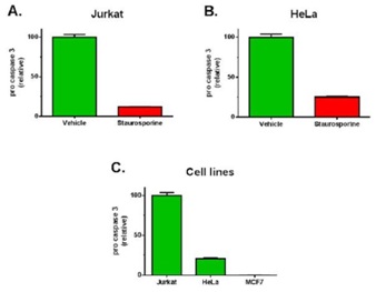
Demonstration of the assay specificity by inducing pro caspase 3 cleavage (A and B). Cells were treated for 4 hours with 1 µM staurosporine or drug’s vehicle (DMSO) and 250 µg/mL Jurkat cell extracts or 1000 µg/mL of HeLa cell extracts were analyzed using this kit. Relative pro caspase 3 levels expressed in percent of vehicle-treated control are shown in A and B. Demonstration of the assay specificity by analyzing cells with variable expression pro caspase 3 (C). Relative pro caspase 3 levels in various human cell lines expressed as percent of Jurkat cells are shown. Note that no pro caspase 3 was detected in MCF7 cells known for their absence of pro caspase 3. Relative pro caspase 3 levels were interpolated from standard curve of Jurkat cell extract dilutions.

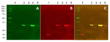
HeLa cells were treated with drugs’ vehicle (DMSO, lane 2), or 1 µM staurosporine (lane 3) for 4 hours. Jurkat cells were treated with drugs’ vehicle (DMSO, lane 4), or 1 µM staurosporine (lane 5) for 4 hours. 20 ng/lane of Active Caspase 3 protein (ab52314, lane 1) and 20 µg/ lane of cell extracts were analyzed by Western blotting using the pro caspase 3 capture antibody (A), the caspase 3 detector antibody (B) and IRDyes labeled secondary antibodies. The overlay of the green signal of the pro caspase 3 capture antibody with the red signal of the caspase 3 detector antibody is shown in C. Note that the pro caspase 3 antibody used as the capture in this kit specifically detects only the pro caspase 3 but not the p17 subunit of the active caspase 3.


