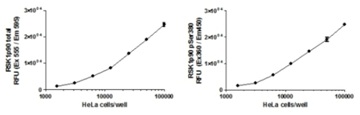
HeLa cells were seeded on amine coated plates within the working range of the assay the day before fixation. Levels of total RSK1 p90 and phosphorylated protein at Ser380 were measured in PMA treated and untreated cells.
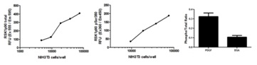
Levels of total RSK1 p90 and phosphorylated protein at Ser380 were measured in serum-starved NIH3T3 cells that were treated with PDGF recombinant protein at 50 ng/mL in 1% BSA. The relative levels of Total RSK1 p90 (left) and RSK1 p90 pSer380 (middle) are shown after background subtraction. Specific phosphorylation was calculated by determining the ratio of pSer380 to Total RSK1 p90 levels after normalization (right).
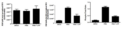
Levels of total RSK1 p90 and phosphorylated protein at Ser380 were measured in DMSO and PMA treated cells. HeLa cells were seeded on amine coated plates within the working range of the assay and serum starved overnight prior to 1 hour treatment with 200 nM PMA or DMSO. Specificity of phosphorylation was determined by treating PMA-treated HeLa cells with LPP at 4,000 units/mL for 45 minutes at 40°C immediately after permeabilization. The relative levels of Total RSK1 p90 (Left) and RSK1 p90 pSer380 (Middle) are shown after background subtraction and normalization with Janus green. Specific Phosphorylation was calculated by determining the ratio of normalized pSer380 to normalized Total RSK1 p90 (Right).
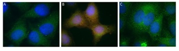
HeLa cells were seeded on glass coverslips and allowed to adhere for a few hours. Cells were then serum starved overnight and treated the next day with DMSO (A) or 200 nM PMA (B and C). Levels of RSK1 p90 total and phosphorylated protein at Ser380 were measured following this protocol. The specificity of signal was determined by treating PMA-treated cells with 4,000 units/mL LPP prior to the assay (C). The total RSK1 p90 antibody was labeled with GAM Alexa 488 (green) whereas the RSK1 p90 pSer380 antibody was labeled with GAR Alexa 594 (red). The nucleus was counterstained with DAPI. The panels show up-regulation of phosphorylation levels due to PMA treatment and overlap of the total and phospho signal shown by the yellow color (B) that is not seen in the DMSO treated cells (A). The PMA induced phosphorylation was removed after treatment with LPP (C).
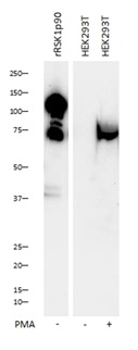
Western blot was run on a 4-20% gradient acrylamide gel. Gel was loaded from left to right: (1) 100 ng of recombinant RSK1p90 (rRSK1p90) ab60880, (2) 40 µg of HEK293T cells treated with DMSO only or (3) HEK 293T cells treated with 200 nM PMA (right).Blocking and secondary antibody incubation steps done in 5% milk, 20 mM Tris-HCl, 0.1% TWEEN-20.Primary incubation steps done in 1% milk, 20 mM Tris-HCL, 0.1% TWEEN-20.
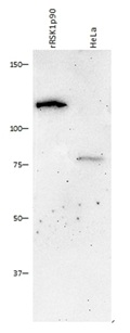
Western blot was run on an 8% acrylamide gel. Samples were loaded from left to right: (1) 100 ng of recombinant RSK1p90 (rRSK1p90) ab60880, (2) 40 µg of HeLa lysate.Blocking and secondary antibody incubation steps were done in 5% milk, 20 mM Tris-HCl, 0.1% TWEEN-20.Primary incubation steps done in 1% milk, 20mM Tris-HCL, 0.1% TWEEN-20.