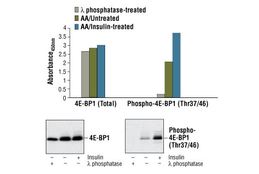
Figure 1: Treatment of HEK-293T cells with amino acids and insulin stimulates phosphorylation of 4E-BP1 at Thr37/46, detected by PathScan ® Phospho-4E-BP1 (Thr37/46) Sandwich ELISA Kit #7216, but does not affect the level of total 4E-BP1 protein detected by PathScan ® Total 4E-BP1 Sandwich ELISA Kit #7179. HEK-293T cells (70-80% confluent) were starved overnight and deprived of amino acids for 1 hour. The amino acids were replenished for 1 hour. Cells were either untreated or stimulated with 100 nM insulin for 30 minutes at 37ºC. λ phosphatase treatment of control cell lysates (4000 U/mL for 60 minutes at 37ºC) abolishes the basal phosphorylation of 4E-BP1 as shown by both sandwich ELISA and Western analysis. The absorbance readings at 450 nm are shown in the top figure while the corresponding Western blots, using 4E-BP1 Antibody #9452 (left panel) or Phospho-4E-BP1 (Thr37/46) Antibody #9459 (right panel), are shown in the bottom figure.

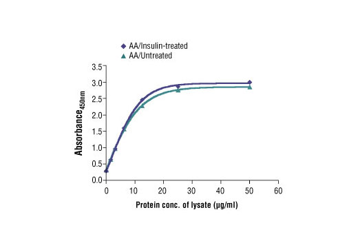
Figure 2: The relationship between the protein concentration of the lysate from amino acid (AA)/untreated and AA/insulin-treated HEK-293T cells and the absorbance at 450 nm is shown.

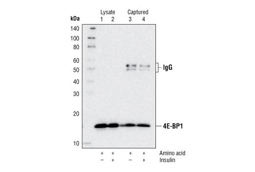
Figure 3. Kit specificity as demonstrated by Western analysis of the ELISA microwell captured protein. Lysates were prepared from HEK-293T cells and incubated in microwells coated with the 4E-BP1 capture antibody. Wells were washed, and the captured protein was solubilized in SDS gel loading buffer. Western analysis of HEK-293T cell starting lysates (lanes 1 & 2) and the captured protein (lanes 3 & 4) was performed using 4E-BP1 Antibody #9452. The major band detected in the captured material (lanes 3 & 4) corresponds to 4E-BP1.


