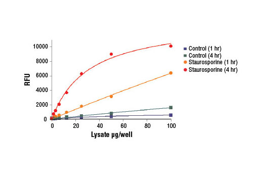
Figure 1. NIH/3T3 cells were treated with Staurosporine #9953 (5 μM, 5 hr) and then lysed in PathScan ® Sandwich ELISA Lysis Buffer (1X) #7018 (supplied with kit). Various amounts of cell lysate were added to assay plates containing the substrate solution, and plates were incubated at 37ºC in the dark. Relative fluorescent units (RFUs) were acquired at 1 and 4 hr.

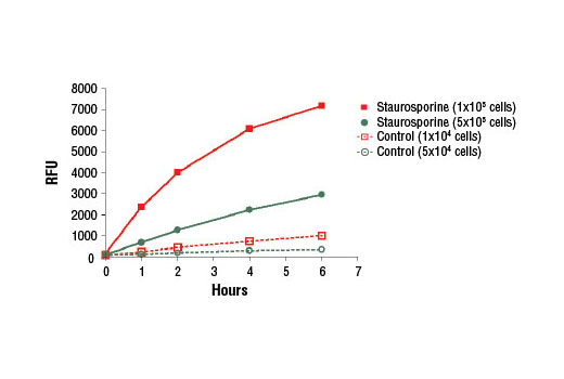
Figure 2. NIH/3T3 cells were seeded in a 96-well plate at 1x10 5 cells/well or 5x10 4 cells/well, and then treated with Staurosporine #9953 (5 μM, 5 hr) and then lysed in 30 μl PathScan ® Sandwich ELISA Lysis Buffer (1X) #7018 (supplied with kit). Cell lysate was added to assay plates containing the substrate solution, and plates were incubated at 37ºC in the dark. Relative fluorescent units (RFUs) were acquired at 0, 1, 2, 4, and 6 hr.

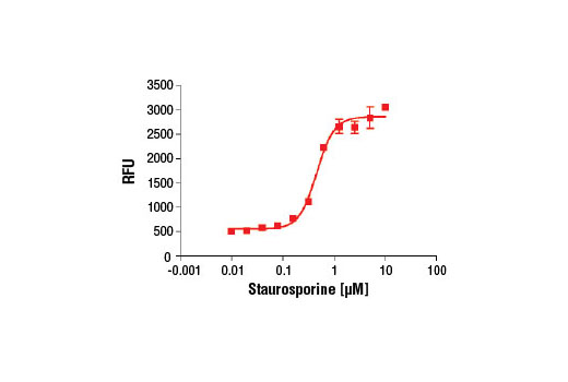
Figure 3. HeLa cells were seeded at 1x10 5 cells/well in a 96-well plate and incubated overnight. Cells were treated with various concentrations of Staurosporine #9953 (5 hr) and then lysed in 30 μL of PathScan ® Sandwich ELISA Lysis Buffer (1X) #7018 (supplied with kit). Cell lysate was mixed with substrate solution and incubated at 37ºC in the dark for 2 hr and relative fluorescent units (RFUs) were acquired.


