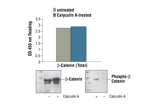
Figure 1: Nonphospho- and phospho-β-catenin proteins from untreated and Calyculin A-treated 293 cells can be detected by PathScan® Total β-Catenin Sandwich ELISA kit #7308 with similar optical density readings. OD 450 readings are shown in the top figure, while the corresponding Western blot using Phospho-β-Catenin (Ser45) Antibody #9564 (right panel) or β-Catenin Antibody #9562 (left panel), is shown in the bottom figure.

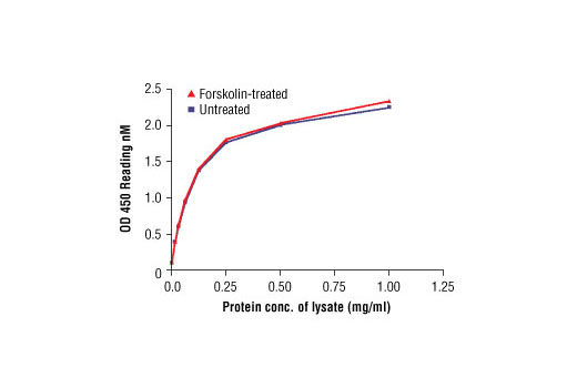
Figure 2: The relationship between protein concentration of lysates from untreated and Forskolin treated COS cells and kit assay optical density readings is shown. COS cells were treated with Forskolin #3828 (50 µM) for 30 min at 37°C, and then lysed.

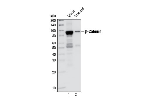
Figure 3: Kit specificity demonstrated by Western blot analysis of the ELISA-well captured protein. Lysates were prepared from human 293 cells and incubated in wells coated with capture β-Catenin Antibody #9562. Wells were then washed and captured protein was solubilized in SDS gel loading buffer. Western blot analysis of 293 cell starting lysate (lane 1) and captured protein (lane 2) was performed, using β-Catenin Mouse mAb #2322. The major band detected in the captured material corresponds to the β-catenin protein (lane 2).


