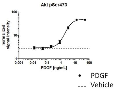
Figure 1. Normalized IR signal in PDGF induced NIH3T3 cells. NIH3T3 cells were seeded at 40,000 cells per well and allowed to adhere for 2 hours prior to serum starvation and induction of phosphorylation with a dose-response of PDGF recombinant protein. At maximum doses, PDGF induced a 5 fold increase in the levels of Akt-1 phosphorylation.
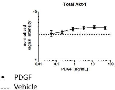
Figure 2. Normalized IR signal in PDGF induced NIH3T3 cells. NIH3T3 cells were seeded at 40,000 cells per well and allowed to adhere for 2 hours prior to serum starvation and induction of phosphorylation with a dose-response of PDGF recombinant protein. At maximum doses, 1.2 fold induction of Akt-1 is much less than that of the Akt-1 phosphorylation (Figure 1).
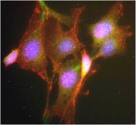
Figure 3. Antibody specificity demonstrated by immunocytochemistry. ICC was carried out on PDGF treated NIH3T3 cells with anti-Akt1 phosphoS473 (ab81283) and anti-Akt1 (ab54752) and all buffer reagents as supplied in this kit. Labeling was carried out with a polyclonal antibody GAR-594 and GAM-488 respectively. The PDGF induced cells show significant induction of Akt phosphorylation at residue S473 (observed in the 594 channel).
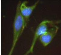
Figure 4. Antibody specificity demonstrated by immunocytochemistry. ICC was carried out on vehicle treated NIH3T3 cells with anti-Akt1 phosphoS473 (ab81283)and anti-Akt1 (ab54752) and all buffer reagents as supplied in this kit. Labeling was carried out with a polyclonal antibody GAR-594 and GAM-488 respectively. This non-induced control shows less induction than compared to the PDGF induced cells showing a significant induction of Akt phosphorylation at residue S473 (figure 3).
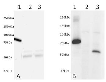
Figure 5. Validation of antibodies by Western Blot. Western blot was run on a 10-20% gradient acrylamide gel. Samples were loaded as follows from left to right: (1) 50ng of Human recombinant AKT1 protein (tagged) (ab62279), (2) 25ug of non-induced NIH3T3 cell extract and (3) 25ug of PDGF induced NIH3T3 cell extract. Membrane Blocking was carried out with 5% Milk+50mM Tris+0.05% Tween-20 pH 7.4, primary antibodies (ab54752 at 5ug/mL left and ab81283 at 1:5000 right) were incubated overnight in 5% BSA+50mM+0.05% Tween-20 pH 7.4 and secondary antibodies were incubated for 2 hours in 5% Milk+50mM Tris+0.05% Tween-20 pH 7.4.