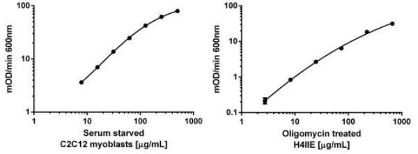
AMPK α1 ELISA assay - dynamic range in rodent cell lines. Relative levels of AMPK α pT172 can be measured in rodent samples. Left panel shows mOD/min data obtained with C2C12 (mouse myoblasts) serum starved overnight in 5mM glucose DMEM media. Right panel shows H4IIE (rat hepatocarcinoma) serum starved overnight in 5mM glucose DMEM media and treated with 1µM Oligomycin for 3 hours.

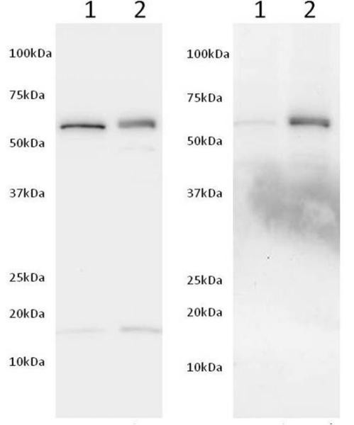
Western blot using capture anti-AMPK α (left) and detector anti-AMKPK α pT172 (right) antibody. The detector antibody used in this kit specifically detects the phosphorylated AMPK α as determined by Western blotting. Hek293T extracts were treated with 1:100 dilution of λ Ppase at 34˚C (lane 1) or untreated (lane 2). Samples were then diluted in SDS-PAGE buffer and loaded at 40 µg/well. Membranes were blocked with 2X blocking buffer (ab126587) diluted in PBS for 1 hour and incubated with either the capture antibody against total AMPK α (Left) or the detector antibody AMPK α phospho T172 (Right) in 1X blocking buffer (ab126587) diluted in PBS/0.05% Tween-20 overnight. Labeling was carried out with secondary antibodies conjugated to HRP. λ Ppase completely dephosphorylates AMPK α without affecting the protein levels.

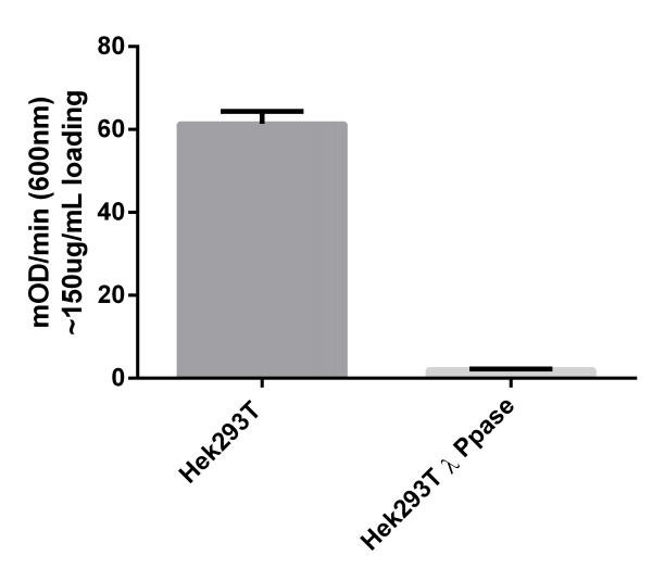
The AMPK α pT172 ELISA specifically measures the phosphorylated threonine. Hek293T extracts were left untreated (control), treated with with 1:100 dilution of λ Ppase at 34˚C. Samples were loaded at 150 µg/mL on the plate and measured following the kit’s protocol. Treatment of Hek293T extracts with λ Ppase decreases the signal to background levels.

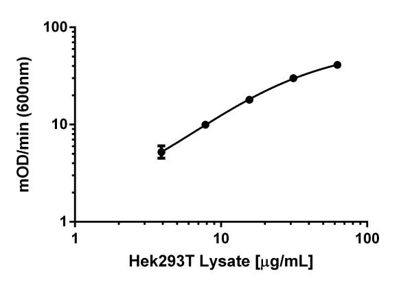
Example of positive control sample standard curve. A dilution series of extract in Incubation Buffer in the working range of the assay. The extract was prepared with Hek293T cells grown in High Glucose DMEM supplemented with 10% FCS.


