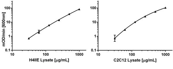
Relative levels of AMPK α1 can be measured in rodent samples. Left panel shows mOD/min data obtained with H4IIE (rat hepatocarcinoma) cells loaded in a range from 60 to 1000 µg/mL. Right panel shows same type of data on C2C12 (mouse myoblasts).
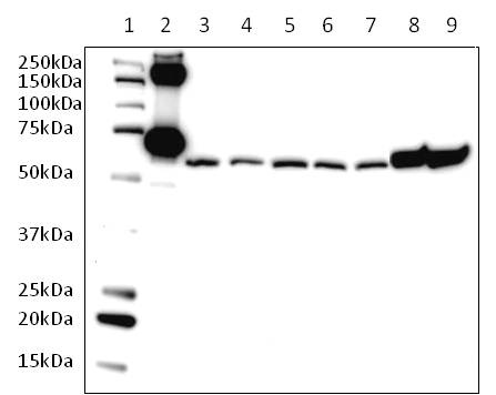
Endogenous levels of AMPK α were measured from various cell lysates on western blot using the capture antibody provided in the kit. Samples were loaded as follows: (1) Marker, (2) AMPK α1 standard protein (MYC/DDK tagged) – 40 ng, (3) C2C12 myotubes – 40 µg (4) C2C12 myoblasts in 10F media – 40 µg, (5) C2C12 myoblasts in 0F media – 40 µg, (6) H4IIE in 10F media – 40 µg, (7) H4IIE in 0F media – 40 µg, (8) Hek293T in 10F media – 40 µg, (9) Hek293T in 0F media – 40 µg. Blot was performed under reducing conditions. Membranes were blocked with 5% Milk in TBS + 0.1% Tween-20 (TBST). Primary antibody was incubated in 1% Milk in TBST overnight at 1/5000. Secondary antibody (GAR HRP) was incubated in 1X blocking buffer.
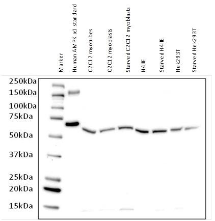
Endogenous levels of AMPK α were measured from various cell lysates on western blot using the capture antibody provided in the kit. Samples were loaded as follows: (1) Marker, (2) AMPK α1 standard protein (MYC/DDK tagged) – 40 ng, (3) C2C12 myotubes – 40 µg (4) C2C12 myoblasts in 10F media – 40 µg, (5) C2C12 myoblasts in 0F media – 40 µg, (6) H4IIE in 10F media – 40 µg, (7) H4IIE in 0F media – 40 µg, (8) Hek293T in 10F media – 40 µg, (9) Hek293T in 0F media – 40 µg. Blot was performed under reducing conditions. Membranes were blocked with 5% Milk in TBS + 0.1% Tween-20 (TBST). Primary antibody was incubated in 1% Milk in TBST overnight at 1/1000. Secondary antibody (GAM HRP) was incubated in 1X blocking buffer.
