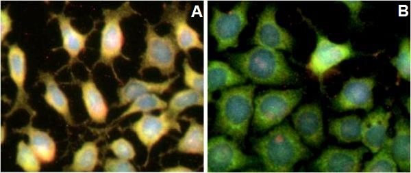
H4IIE cells were seeded on glass coverslips and allowed to adhere for a few hours. Cells were then serum starved overnight and treated the next day with 1 µM oligomycin (left) or DMSO (right). Levels of AMPKα total and phosphorylated protein at T172/183 were measured following this protocol. The total AMPKα signal is shown in green and AMPKα p172 in red. The left panel shows up-regulation of phosphorylation levels due to oligomycin treatment.

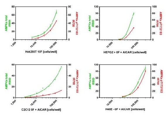
Cells were seeded the day before fixation at the specified cell density and allowed to adhere overnight. Cells were then fixed the next day and signal was obtained using this kit as described. In this experiment, the Hek293T cells were instead permeabilized with methanol at -20°C due to their sensitivity to antigen retrieval. Total AMPKα and AMPKα pT172 are shown after background subtraction.

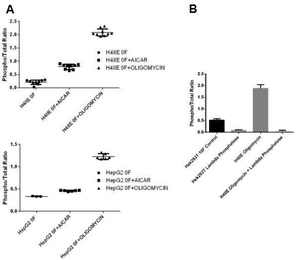
(A) H4IIE and HepG2 cells were seeded on amine coated plates within the working range of the assay the day before fixation. Levels of total AMPKα and phosphorylated protein at T172/183 were measured after serum starvation and treatment with AICAR (ab120358) or oligomycin. Normalized signal intensities were ratio to show the effect of treatment on the phosphorylation status of AMPK. (B) H4IIE cells treated with oligomycin and untreated Hek293T were fixed on a 96 well plates at densities within the working range of the assay. After fixation, cells were permeabilized with methanol at -20°C for 30 minutes and treated with and without Lamdba Phosphatase at 40˚C for 45 minutes on a plate heater. Blocking and antibody incubations were carried out according to this protocol (without the use of Triton X-100). Normalized signal intensities were ratio to show the effect of treatment on the phosphorylation status of AMPK.

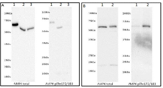
Western Blot was run on a 4-20% gradient acrylamide gel. (A) Samples were loaded as follows: (1) 40ng of AMPKα human recombinant protein, (2) 40 µg of C2C12 myoblasts serum starved (3) 40 µg of C2C12 myoblasts in 10% FCS. (B) Samples were loaded as follows: (1) 40 µg Hek293T in 10% FCS treated with 1/100 dilution of LP (2) 40 µg Hek293T in 10% FCS. AMPKα total membrane was blocked with 5% Milk in TBST, AMPKα pT172 was blocked with 1X Blocking buffer (ab126587) in TBST.


