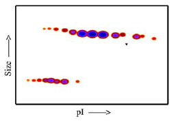
The densitometry scan demonstrates the purified human cell expressed protein exists in multiple isoforms, which differ according to their level of post-translational modification. The triangle indicates theoretical pI and MW of the protein.

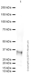
Anti-VEGFC antibody ( ab83905 ) at 1 µg/ml + Human VEGFC full length protein (ab83573) at 0.1 µg Secondary Goat Anti-Rabbit IgG H&L (HRP) preadsorbed ( ab97080 ) at 1/5000 dilution developed using the ECL technique Performed under reducing conditions. Predicted band size : 46 kDa Exposure time : 4 minutes

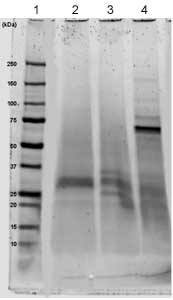
Lane 1 – MW markers; Lane 2 – ab83573 ; Lane 3 – ab83573 treated with PNGase F to remove potential N-linked glycans; Lane 4 – ab83573 treated with a glycosidase cocktail to remove potential N- and O-linked glycans. Approximately 5 µg of protein was loaded per lane. Drop in MW after treatment with PNGase F indicates presence of N-linked glycans. A possible further drop in MW after treatment with the glycosidase cocktail indicates the possible presence of O-linked glycans. Additional bands in lane 3 and lane 4 are glycosidase enzymes.

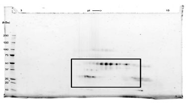
2D SDS-PAGE: A sample of VEGFC without carrier protein was reduced and alkylated and focused on a 3-10 IPG strip then run on a 4-20% Tris-HCl 2D gel. Approximately 40 µg of protein was loaded. Spot train indicates presence of multiple isoforms of VEGFC. Spots within the spot train were cut from the gel and identified as VEGFC by protein mass fingerprinting.


