 Immunohistochemistry
Rabbit Anti-HDAC1 Antibody, Affinity Purified:
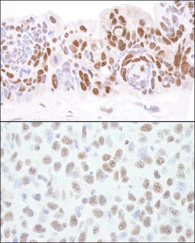
Cat. No. PLA0134: Detection of Human and Mouse HDAC1 by Immunohistochemistry. Sample: FFPE section of Human ovarian carcinoma (top) and Mouse squamousl cell carcinoma (bottom). Antibody: Affinity purified Rabbit Anti-HDAC1 (
Cat. No. PLA0134) used at a dilution of 1:200 (1 μg/mL) and 1:1,000 (0.2 μg/mL). Detection: DAB
 Immunofluorescence
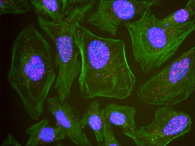
: This antibody is qualified for the Proximity Ligation Assay (PLA). The above image shows representative results for PLA using three color fluorescence, including DAPI stained nuclei (blue), phalloidin stained cytoplasmic F-actin (green), and PLA positive signal (red).
 Immunoblotting
Rabbit Anti-HDAC1 Antibody, Affinity Purified:
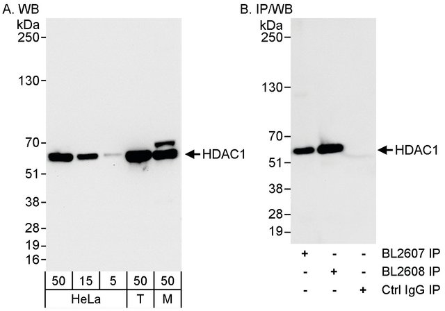
Cat. No. PLA0134: Detection of Human and Mouse HDAC1 by WB and Immunoprecipitation. Samples: Whole cell lysate from HeLa (5, 15 and 50 μg for WB; 1 mg for IP, 20% of IP loaded), 293T (Lane T; 50 μg), and Mouse NIH3T3 (Lane M; 50 μg) cells. Antibodies: Affinity purified Rabbit Anti-HDAC1 Antibody (
Cat. No. PLA0134) used for WB at 0.04 μg/mL (A) and 1 μg/mL (B) and used for IP at 3 μg/mg lysate (B). HDAC1 was also immunoprecipitated using a Rabbit Anti-HDAC1 Antibody recognizing a different epitope at 3 μg/mg lysate. Detection: Chemiluminescence with exposure times of 30 seconds (A) and 10 seconds (B).


