MAP3K7

Gene Symbol MAP3K7
Entrez Gene 6885
Alt Symbol MEKK7, TAK1, TGF1a
Species Human
Gene Type protein-coding
Description mitogen-activated protein kinase kinase kinase 7
Other Description TGF-beta activated kinase 1|TGF-beta-activated kinase 1|transforming growth factor-beta-activated kinase 1
Swissprots B2RE27 O43317 Q5TDT7 Q5TDN2 Q9NZ70 O43318 O43319 E1P523 Q5TDN3 Q9NTR3
Accessions ABC40734 EAW48525 EAW48526 EAW48527 EAW48528 EAW48529 O43318 AB009356 BAA25025 AB009357 BAA25026 AB009358 BAA25027 AF218074 AAF27652 AI147171 AK055901 AK299268 BAG61290 AK315774 BAG38124 AL050393 CAB43687 AL520975 BC017715 AAH17715 BM172351 BT019654 AAV38460 BT019655 AAV38461 BX648277 XM_006715553 XP_006715616 NM_003188 NP_003179 NM_145331 NP_663304 NM_145332 NP_663305 NM_145333 NP_663306
Function Serine/threonine kinase which acts as an essential component of the MAP kinase signal transduction pathway. Plays an important role in the cascades of cellular responses evoked by changes in the environment. Mediates signal transduction of TRAF6, various cytokines including interleukin-1 (IL-1), transforming growth factor-beta (TGFB), TGFB-related factors like BMP2 and BMP4, toll-like receptors (TLR), tumor necrosis factor receptor CD40 and B-cell receptor (BCR). Ceramides are also able to activate MAP3K7/TAK1. Once activated, acts as an upstream activator of the MKK/JNK signal transduction cascade and the p38 MAPK signal transduction cascade through the phosphorylation and activation of several MAP kinase kinases like MAP2K1/MEK1, MAP2K3/MKK3, MAP2K6/MKK6 and MAP2K7/MKK7. These MAP2Ks in turn activate p38 MAPKs, c-jun N-terminal kinases (JNKs) and I-kappa-B kinase complex (IKK). Both p38 MAPK and JNK pathways control the transcription factors activator protein-1 (AP-1), while nuclear
Subcellular Location Cytoplasm {ECO:0000269|PubMed:12242293}. Cell membrane {ECO:0000269|PubMed:12242293}; Peripheral membrane protein {ECO:0000269|PubMed:12242293}; Cytoplasmic side {ECO:0000269|PubMed:12242293}. Note=Although the majority of MAP3K7/TAK1 is found in the cytosol, when complexed with TAB1/MAP3K7IP1 and TAB2/MAP3K7IP2, it is also localized at the cell membrane.
Tissue Specificity Isoform 1A is the most abundant in ovary, skeletal muscle, spleen and blood mononuclear cells. Isoform 1B is highly expressed in brain, kidney and small intestine. Isoform 1C is the major form in prostate. Isoform 1D is the less abundant form. {ECO:0000269|PubMed:11118615}.
Top Pathways AMPK signaling pathway, Epstein-Barr virus infection, Herpes simplex infection, TNF signaling pathway, Wnt signaling pathway

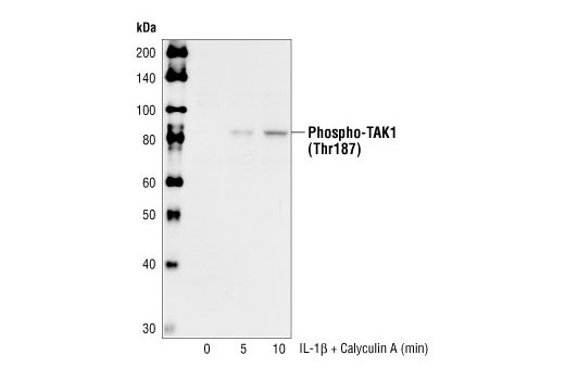
Phospho-TAK1 (Thr187) Antibody - 4536 from Cell Signaling Technology

Prices $287.00
Sizes 100 µl (10 western blots)
Host Rabbit
Clonality Polyclonal
Western blot analysis of extracts from 293IL-1R cells, untreated or treated with IL-1beta and Calyculin A for the indicated times, using Phospho-TAK1 (Thr187) Antibody.

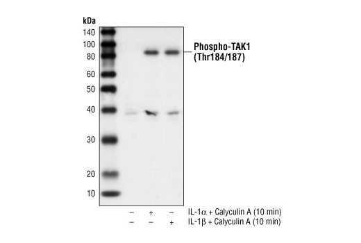
Phospho-TAK1 (Thr184/187) Antibody - 4531 from Cell Signaling Technology

Prices $287.00
Sizes 100 µl (10 western blots)
Host Rabbit
Clonality Polyclonal
Western blot analysis of extracts from 293 IL-1R cells treated with IL-1alpha (20 ng/ml) or IL-1beta (10 ng/ml) and Calyculin A (50 nM) for 10 minutes, using Phospho-TAK1 (Thr184/187) Antibody.

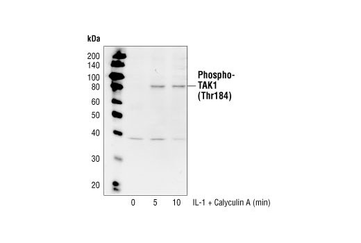
Phospho-TAK1 (Thr184) Antibody - 4537 from Cell Signaling Technology

Prices $287.00
Sizes 100 µl (10 western blots)
Host Rabbit
Clonality Polyclonal
Western blot analysis of extracts from 293 IL-1R cells treated with IL-1beta (10 ng/ml) and Calyculin A (50 nM) for the indicated times, using Phospho-TAK1 (Thr184) Antibody.

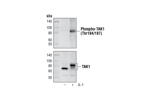
Phospho-TAK1 (Thr184/187) (90C7) Rabbit mAb - 4508 from Cell Signaling Technology

Prices $287.00, $678.00
Sizes 100 µl (10 western blots), 300 µl (30 western blots)
Host Rabbit
Clonality Monoclonal
Clone 90C7
Western blot analysis of extracts from 293 IL-1R cells, untreated or treated for 10 minutes with IL-1, using Phospho-TAK1 (Thr184/187) (90C7) Rabbit mAb (upper) or TAK1 Antibody #4505 (lower).

Phospho-TAK1 (Ser412) Antibody - 9339 from Cell Signaling Technology

Prices $287.00
Sizes 100 µl (10 western blots)
Host Rabbit
Clonality Polyclonal
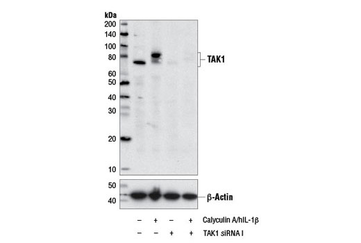
Western blot analysis of extracts from HeLa cells co-treated with and without 100nM Calyculin A (#9902) and 20 ng/mL hIL-1β (#8900) for 10 min., and transfected with and without 100nM SignalSilence ® TAK1 siRNA I (#6317) using Phospho-TAK1 (Ser412) Antibody #9339 (top) and β-actin (13E5) Rabbit mAb #4970 (bottom).

TAK1 Antibody - 4505 from Cell Signaling Technology

Prices $246.00
Sizes 100 µl (10 western blots)
Host Rabbit
Clonality Polyclonal
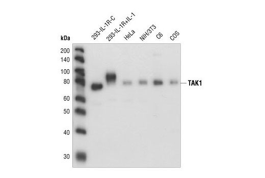
Western blot analysis of extracts from untreated or IL-1 treated 293-IL-1R cells, and extracts from HeLa, NIH/3T3, C6, and COS cells, using TAK1 Antibody.

TAK1 (D94D7) Rabbit mAb - 5206 from Cell Signaling Technology

Prices $246.00
Sizes 100 µl (10 western blots)
Host Rabbit
Clonality Monoclonal
Clone D94D7
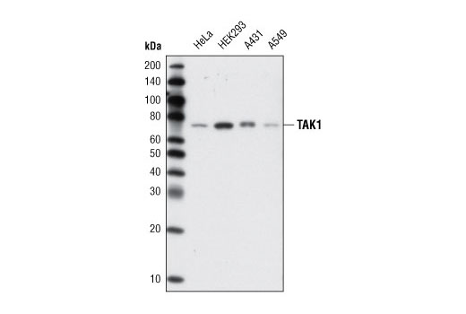
Western blot analysis of extracts from various cell lines using TAK1 (D94D7) Rabbit mAb.

Tak1 (C-9) - sc-7967 from Santa Cruz Biotechnology

Prices $279.00
Sizes 200 µg/ml
Host Mouse
Clonality Monoclonal
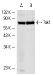
Tak1 (C-9): sc-7967. Western blot analysis of Tak1 expression in HeLa (A) and 3611-RF (B) whole cell lysates.

Tak1 (M-17) - sc-1839 from Santa Cruz Biotechnology

Prices $279.00
Sizes 200 µg/ml
Host Goat
Clonality Polyclonal
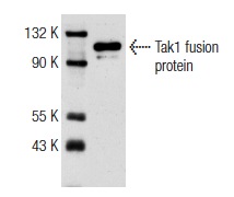
Tak1 (M-17): sc-1839. Western blot analysis of human recombinant Tak1 fusion protein.

Tak1 (H-5) - sc-166562 from Santa Cruz Biotechnology

Prices $279.00
Sizes 200 µg/ml
Host Mouse
Clonality Monoclonal
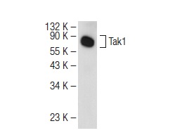
Tak1 (H-5): sc-166562. Western blot analysis of Tak1 expression in 3611-RF whole cell lysate.

Tak1 (M-579) - sc-7162 from Santa Cruz Biotechnology

Prices $279.00
Sizes 200 µg/ml
Host Rabbit
Clonality Polyclonal
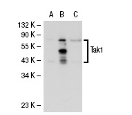
Tak1 (M-579): sc-7162. Western blot analysis of Tak1 expression in non-transfected: sc-110760 (A) and human Tak1 transfected: sc-113194 (B) 293 whole cell lysates.

p-Tak1 (Ser 192) - sc-130219 from Santa Cruz Biotechnology

Prices $279.00
Sizes 100 µg/ml
Host Rabbit
Clonality Polyclonal

Anti-TAK1 (phospho S439) antibody [EPR2863] - ab109404 from Abcam

Prices $357.00
Sizes 100 µl
Host Rabbit
Clonality Monoclonal
Clone EPR2863
All lanes : Anti-TAK1 (phospho S439) antibody [EPR2863] (ab109404) at 1/1000 dilutionLane 1 : HeLa cell lysateLane 2 : HeLa cell lysate treated with transforming growth factor-beta (TGF-beta)Lysates/proteins at 10 µg per lane.

Anti-TAK1 (phospho T187) antibody - ab192443 from Abcam

Prices $370.00
Sizes 100 µg
Host Rabbit
Clonality Polyclonal
All lanes : Anti-TAK1 (phospho T187) antibody (ab192443) at 1/500 dilutionLane 1 : NIH-3T3 whole cell lysate with antigen-specific peptideLane 2 : NIH-3T3 whole cell lysateLysates/proteins at 35 µg per lane.SecondaryPeroxidase-conjugated goat anti-rabbit IgG (H+L) at 1/5000 dilution

Anti-TAK1 antibody - ab25879 from Abcam

Prices $378.00
Sizes 100 µg
Host Rabbit
Clonality Polyclonal
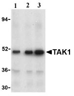
Lane 1 :  Anti-TAK1 antibody (ab25879) at 1 µg/mlLane 2 :  Anti-TAK1 antibody (ab25879) at 2 µg/mlLane 3 :  Anti-TAK1 antibody (ab25879) at 4 µg/mlLane 1 : Rat Thymus Tissue Lysate (ab4041)Lane 2 : Rat Thymus Tissue Lysate (ab4041)Lane 3 : Rat Thymus Tissue Lysate (ab4041)

Anti-TAK1 antibody - ab79363 from Abcam

Prices $378.00
Sizes 100 µg
Host Rabbit
Clonality Polyclonal
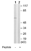
All lanes : Anti-TAK1 antibody (ab79363) at 1/500 dilutionLane 1 : HeLa cell extractLane 2 : HeLa cell extract with immunising peptide at 10 µgLysates/proteins at 30 µg per lane.

Anti-TAK1 antibody - ab196955 from Abcam

Prices $376.00
Sizes 100 µl
Host Rabbit
Clonality Polyclonal
Immunohistochemical analysis of paraffin-embedded Human breast carcinoma tissue labeling TAK1 with ab196955 at 1/50 dilution. The image on the right is preincubated with blocking peptide.

Anti-TAK1 antibody - ab63252 from Abcam

Prices $376.00
Sizes 100 µg
Host Rabbit
Clonality Polyclonal
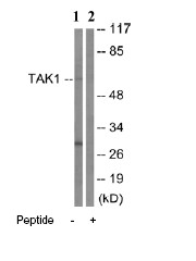
All lanes : Anti-TAK1 antibody (ab63252) at 1/500 dilutionLane 1 : extracts from HepG2 cellsLane 2 : extracts from HepG2 cells with immunising peptide at 10 µgLysates/proteins at 30 µg per lane.

Anti-TAK1 antibody [EPR5984] - ab109526 from Abcam

Prices $381.00
Sizes 100 µl
Host Rabbit
Clonality Monoclonal
Clone EPR5984
All lanes : Anti-TAK1 antibody [EPR5984] (ab109526) at 1/1000 dilutionLane 1 : K562 cell lysateLane 2 : HeLa cell lysateLane 3 : A431 cell lysateLysates/proteins at 10 µg per lane.SecondaryHRP labelled Goat anti-Rabbit IgG at 1/2000 dilution

Anti-TAK1 antibody [EPR5984] (Alexa Fluor® 488) - ab199191 from Abcam

Prices $425.00
Sizes 100 µl
Host Rabbit
Clonality Monoclonal
Clone EPR5984
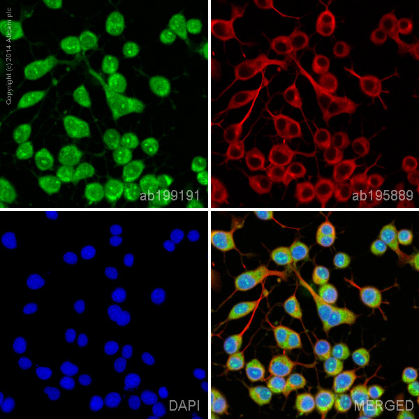
ab199191 staining TAK1 in differentiated Neuro2a cells. The cells were fixed with 100% methanol (5 min), permeabilized with 0.1% Triton X-100 for 5 minutes and then blocked with 1% BSA/10% normal goat serum/0.3M glycine in 0.1% PBS-Tween for 1h. The cells were then incubated overnight at +4°C with ab199191 at a 1/100 dilution (shown in green) and ab195889, Mouse monoclonal to alpha Tubulin (Alexa Fluor® 594), at a 1/250 dilution (shown in red). Nuclear DNA was labelled with DAPI (shown in blue).Image was taken with a confocal microscope (Leica-Microsystems, TCS SP8).

Anti-TAK1 antibody [EPR5984] (Alexa Fluor® 647) - ab199473 from Abcam

Prices $425.00
Sizes 100 µl
Host Rabbit
Clonality Monoclonal
Clone EPR5984
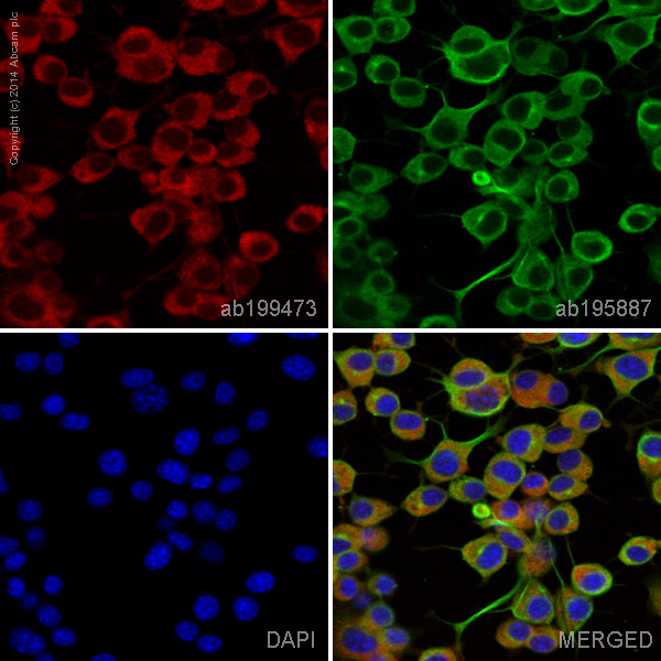
ab199473 staining TAK1 in differentiated Neuro2a cells. The cells were fixed with 100% methanol (5 min), permeabilized with 0.1% Triton X-100 for 5 minutes and then blocked with 1% BSA/10% normal goat serum/0.3M glycine in 0.1% PBS-Tween for 1h. The cells were then incubated overnight at +4°C with ab199473 at a 1/100 dilution (shown in red) and ab195887, Mouse monoclonal to alpha Tubulin (Alexa Fluor® 488), at a 1/250 dilution (shown in green). Nuclear DNA was labelled with DAPI (shown in blue).Image was taken with a confocal microscope (Leica-Microsystems, TCS SP8).This product also gave a positive signal under the same testing conditions in differentiated Neuro2a cells fixed with 4% formaldehyde (10 min).

Anti-TAK1 antibody - N-terminal - ab171757 from Abcam

Prices $376.00
Sizes 400 µl
Host Rabbit
Clonality Polyclonal
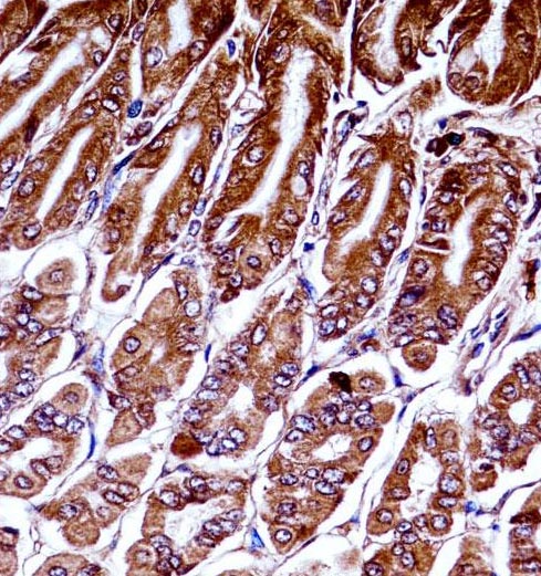
Immunohistochemical analysis of formalin fixed and paraffin embedded mouse stomach tissue labeling TAK1 with ab171757 at 1/10 dilution followed by peroxidase conjugation of the secondary antibody and DAB staining.

Anti-MAP3K7 / TAK1 Antibody (aa563-579) IHC-plus™ - LS-B3754 from LifeSpan Bioscience

Prices $395.00
Sizes 50 µg
Host Rabbit
Clonality Polyclonal
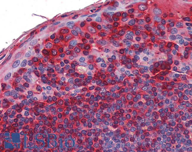
Anti-MAP3K7 / TAK1 antibody IHC of human tonsil. Immunohistochemistry of formalin-fixed, paraffin-embedded tissue after heat-induced antigen retrieval. Antibody LS-B3754 concentration 5 ug/ml.

Anti-MAP3K7 / TAK1 Antibody (aa525-537) - LS-C55561 from LifeSpan Bioscience

Prices $395.00
Sizes 100 µg
Host Goat
Clonality Polyclonal
Antibody (1 ug/ml) staining of HeLa cell lysate (35 ug protein in RIPA buffer). Primary incubation was 1 hour. Detected by chemiluminescence.

Anti-MAP3K7 / TAK1 Antibody (phospho-Thr187) - LS-C97030 from LifeSpan Bioscience

Prices $295.00
Sizes 400 µl
Host Rabbit
Clonality Polyclonal
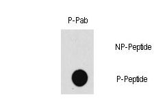
Dot blot of Phospho-MAP3K7-T187 polyclonal antibody on nitrocellulose membrane. 50ng of Phospho-peptide or Non Phospho-peptide per dot were adsorbed. Antibody working concentration were 0.5ug per ml.
Search more