| Prices |
$299.00 |
| Sizes |
100 µl (50 tests) |
| Host |
Rabbit |
| Clonality |
Monoclonal |
| Clone |
D3B5 |
|
| Prices |
$299.00 |
| Sizes |
100 µl (50 tests) |
| Host |
Rabbit |
| Clonality |
Monoclonal |
| Clone |
D3B5 |
|
| Prices |
$299.00 |
| Sizes |
100 µl (50 tests) |
| Host |
Rabbit |
| Clonality |
Monoclonal |
| Clone |
D3B5 |
|
|
|
| Prices |
$99.00, $246.00 |
| Sizes |
20 µl (80 sections), 100 µl (400 sections) |
| Host |
Rabbit |
| Clonality |
Monoclonal |
| Clone |
D3B5 |
|
| Prices |
$99.00, $246.00 |
| Sizes |
20 µl (80 tests), 100 µl (400 tests) |
| Host |
Rabbit |
| Clonality |
Monoclonal |
| Clone |
D3B5 |
|
| Prices |
$99.00, $246.00 |
| Sizes |
20 µl (80 sections), 100 µl (400 sections) |
| Host |
Mouse |
| Clonality |
Monoclonal |
| Clone |
8D5 |
|
| Prices |
$99.00, $246.00 |
| Sizes |
20 µl (80 sections), 100 µl (400 sections) |
| Host |
Rabbit |
| Clonality |
Monoclonal |
| Clone |
D2H10 |
|
| Prices |
$279.00 |
| Sizes |
200 µg/ml |
| Host |
Goat |
| Clonality |
Polyclonal |
|
|
|
| Prices |
$279.00 |
| Sizes |
200 µg/ml |
| Host |
Rabbit |
| Clonality |
Polyclonal |
|
| Prices |
$279.00 |
| Sizes |
200 µg/ml |
| Host |
Mouse |
| Clonality |
Monoclonal |
|
| Prices |
$279.00 |
| Sizes |
200 µg/ml |
| Host |
Goat |
| Clonality |
Polyclonal |
|
| Prices |
$279.00 |
| Sizes |
250 µl supernatant |
| Host |
Mouse |
| Clonality |
Monoclonal |
|
| Prices |
$279.00 |
| Sizes |
1000 µl supernatant |
| Host |
Mouse |
| Clonality |
Monoclonal |
|
|
|
| Prices |
$401.00 |
| Sizes |
200 µl |
| Host |
Rabbit |
| Clonality |
Polyclonal |
|
| Prices |
$394.00 |
| Sizes |
500 µl |
| Host |
Rabbit |
| Clonality |
Polyclonal |
|
| Prices |
$378.00 |
| Sizes |
100 µg |
| Host |
Rabbit |
| Clonality |
Polyclonal |
|
| Prices |
$370.00 |
| Sizes |
200 µl |
| Host |
Goat |
| Clonality |
Polyclonal |
|
| Host |
Mouse |
| Clonality |
Monoclonal |
| Clone |
4A1 |
|
|
|
| Prices |
$376.00 |
| Sizes |
100 µl |
| Host |
Rat |
| Clonality |
Monoclonal |
| Clone |
5D7 |
|
| Prices |
$395.00 |
| Sizes |
250 µl |
| Host |
Mouse |
| Clonality |
Monoclonal |
| Clone |
B126.1 |
|
| Prices |
$378.00 |
| Sizes |
250 µl |
| Host |
Rabbit |
| Clonality |
Monoclonal |
| Clone |
E18-E |
|
| Prices |
$398.00 |
| Sizes |
100 µl |
| Host |
Rabbit |
| Clonality |
Monoclonal |
| Clone |
EPR3610 |
|
| Prices |
$425.00 |
| Sizes |
100 µl |
| Host |
Rabbit |
| Clonality |
Monoclonal |
| Clone |
EPR3610 |
|
| Prices |
$425.00 |
| Sizes |
100 µl |
| Host |
Rabbit |
| Clonality |
Monoclonal |
| Clone |
EPR3610 |
|
| Prices |
$385.00 |
| Sizes |
100 µl |
| Host |
Rabbit |
| Clonality |
Monoclonal |
| Clone |
EPR3611 |
|


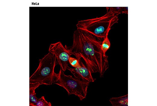

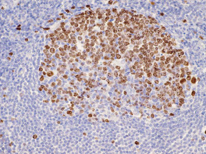
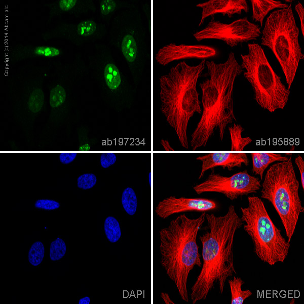
![ab196907 staining Ki67 in HeLa cells. The cells were fixed with 4% formaldehyde (10 min), permeabilized in 0.1% Triton X-100 for 5 minutes and then blocked in 1% BSA/10% normal goat serum/0.3M glycine in 0.1% PBS-Tween for 1h. The cells were then incubated with ab196907 at 1/100 dilution (shown in red) and ab195887, Mouse monoclonal [DM1A] to alpha Tubulin (Alexa Fluor® 488, shown in green) at 1/167 dilution, overnight at +4°C. Nuclear DNA was labelled in blue with DAPI.This product also gave a positive signal under the same testing conditions in HeLa cells fixed with 100% methanol (5 min). Image was taken with a confocal microscope (Leica-Microsystems, TCS SP8)](http://www.bioprodhub.com/system/product_images/ab_products/2/sub_3/12510_ab196907-237107-ICCAP220459502.jpg)