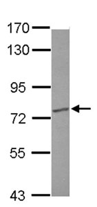
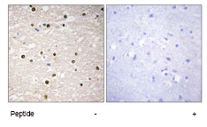
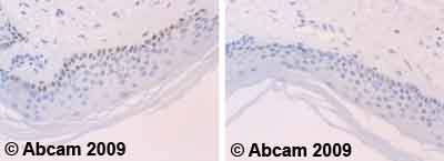
TP73
| Gene Symbol | TP73 |
|---|---|
| Entrez Gene | 7161 |
| Alt Symbol | P73 |
| Species | Human |
| Gene Type | protein-coding |
| Description | tumor protein p73 |
| Other Description | p53-like transcription factor|p53-related protein |
| Swissprots | Q5TBV6 Q17RN8 O15351 Q8NHW9 O15350 B7Z8Z1 C9J521 B7Z9C1 B7Z7J4 Q5TBV5 Q9NTK8 Q8TDY6 Q8TDY5 |
| Accessions | AAC61887 AAD39696 AAL38007 BAB87246 EAW71463 EAW71464 EAW71465 EAW71466 EAW71467 EAW71468 O15350 AB055065 BAB87244 AB055066 BAB87245 AI094238 AK295669 BAH12150 AK302118 BAH13630 AK304177 BAH14127 AK304784 BAH14257 AK310432 AY040827 AAK81884 AY040828 AAK81885 AY040829 AAK81886 BC117251 AAI17252 BC117253 AAI17254 DA439168 DA446656 DC345760 HQ258345 ADR83099 Y11416 CAA72219 XM_011542064 XP_011540366 NM_001126240 NP_001119712 NM_001126241 NP_001119713 NM_001126242 NP_001119714 NM_001204184 NP_001191113 NM_001204185 NP_001191114 NM_001204186 NP_001191115 NM_001204187 NP_001191116 NM_001204188 NP_001191117 NM_001204189 NP_001191118 NM_001204190 NP_001191119 NM_001204191 NP_001191120 NM_001204192 NP_001191121 NM_005427 NP_005418 |
| Function | Participates in the apoptotic response to DNA damage. Isoforms containing the transactivation domain are pro-apoptotic, isoforms lacking the domain are anti-apoptotic and block the function of p53 and transactivating p73 isoforms. May be a tumor suppressor protein. {ECO:0000269|PubMed:10203277, ECO:0000269|PubMed:11753569, ECO:0000269|PubMed:18174154}. |
| Subcellular Location | Nucleus. Cytoplasm. Note=Accumulates in the nucleus in response to DNA damage. |
| Tissue Specificity | Expressed in striatal neurons of patients with Huntington disease (at protein level). Brain, kidney, placenta, colon, heart, liver, spleen, skeletal muscle, prostate, thymus and pancreas. Highly expressed in fetal tissue. {ECO:0000269|PubMed:11753569, ECO:0000269|PubMed:16461361}. |
| Top Pathways | p53 signaling pathway, Neurotrophin signaling pathway, Measles, Hippo signaling pathway |
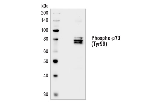
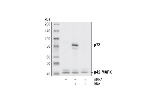
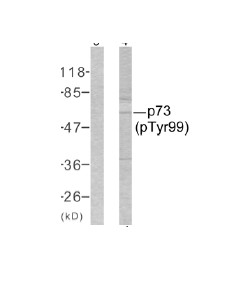
Phospho-p73 (Tyr99) Antibody - 4665 from Cell Signaling Technology
|
||||||||||
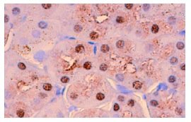
p73 (D3G10) Rabbit mAb - 14620 from Cell Signaling Technology
|
||||||||||
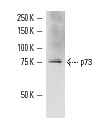
p73 Antibody - 4662 from Cell Signaling Technology
|
||||||||||
ΔN p73 (N-16) - sc-23429 from Santa Cruz Biotechnology
|
||||||||||
ΔN p73 (4H223) - sc-70966 from Santa Cruz Biotechnology
|
||||||||||
ΔN p73 (38C674) - sc-56086 from Santa Cruz Biotechnology
|
||||||||||
p73 (S-20) - sc-9651 from Santa Cruz Biotechnology
|
||||||||||
p73 (N-19) - sc-7236 from Santa Cruz Biotechnology
|
||||||||||
p73 (H-79) - sc-7957 from Santa Cruz Biotechnology
|
||||||||||
p73 (E-4) - sc-17823 from Santa Cruz Biotechnology
|
||||||||||
p73 (C-20) - sc-7237 from Santa Cruz Biotechnology
|
||||||||||
p73 (5B429) - sc-56191 from Santa Cruz Biotechnology
|
||||||||||
p73 (5B1288) - sc-56190 from Santa Cruz Biotechnology
|
||||||||||
p73α (C-17) - sc-7238 from Santa Cruz Biotechnology
|
||||||||||
p73α (SPM431) - sc-56194 from Santa Cruz Biotechnology
|
||||||||||
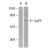
p-p73 (Tyr 99) - sc-101769 from Santa Cruz Biotechnology
|
||||||||||
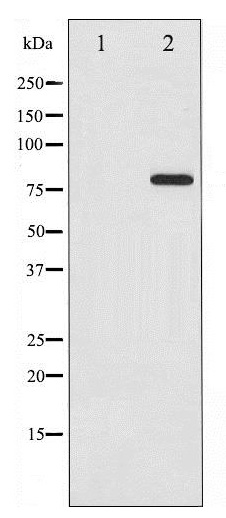
Anti-p73 (phospho Y99) antibody - ab38457 from Abcam
|
||||||||||
Anti-p73 (phospho Y99) antibody - ab195749 from Abcam
|
||||||||||
Anti-p73 alpha+beta antibody - ab31847 from Abcam
|
||||||||||
Anti-p73 alpha+beta antibody [ER-15] - ab17230 from Abcam
|
||||||||||
Anti-p73 alpha+beta antibody [ER-15] - ab80701 from Abcam
|
||||||||||
Anti-p73 antibody - ab137797 from Abcam
|
||||||||||
Anti-p73 antibody - ab94554 from Abcam
|
||||||||||
Anti-p73 antibody - ab70093 from Abcam
|
||||||||||
Anti-p73 antibody - ab39424 from Abcam
|

![Overlay histogram showing HEK293 cells stained with ab17230 (red line). The cells were fixed with 80% methanol (5 min) and then permeabilized with 0.1% PBS-Tween for 20 min. The cells were then incubated in 1x PBS / 10% normal goat serum / 0.3M glycine to block non-specific protein-protein interactions followed by the antibody (ab17230, 1μg/1x106 cells) for 30 min at 22°C. The secondary antibody used was DyLight® 488 goat anti-mouse IgG (H+L) (ab96879) at 1/500 dilution for 30 min at 22°C. Isotype control antibody (black line) was mouse IgG1 [ICIGG1] (ab91353, 1μg/1x106 cells) used under the same conditions. Unlabelled sample (blue line) was also used as a control. Acquisition of >5,000 events were collected using a 20mW Argon ion laser (488nm) and 525/30 bandpass filter. This antibody gave a positive signal in HEK293 cells fixed with 4% paraformaldehyde (10 min)/permeabilized with 0.1% PBS-Tween for 20 min used under the same conditions.](http://www.bioprodhub.com/system/product_images/ab_products/2/sub_4/6162_ab17230-1-ab17230FC.jpg)
