CD55
| Gene Symbol | CD55 |
|---|---|
| Entrez Gene | 1604 |
| Alt Symbol | CR, CROM, DAF, TC |
| Species | Human |
| Gene Type | protein-coding |
| Description | CD55 molecule, decay accelerating factor for complement (Cromer blood group) |
| Other Description | CD55 antigen|complement decay-accelerating factor |
| Swissprots | D3DT84 P78361 Q14UF3 B1AP14 P09679 Q14UF4 D3DT83 Q14UF2 Q14UF5 E7ER69 Q14UF6 P08174 |
| Accessions | AAA52170 AAC60633 AAW29942 BAA22900 CBX47421 EAW93485 EAW93486 EAW93487 EAW93488 EAW93489 EAW93490 EAW93491 EAW93492 EAW93493 EAW93494 EAW93495 EAW93496 EAW93497 EAW93498 EAW93499 P08174 AB240566 BAE97422 AB240567 BAE97423 AB240568 BAE97424 AB240569 BAE97425 AB240570 BAE97426 AF052110 AK298549 BAG60746 AK300621 BAG62314 AK312448 BAG35355 AW316643 AY055757 AAL25832 AY055758 AAL25833 AY055759 AAL25834 AY055760 AAL25835 BC001288 AAH01288 BI545505 BT007159 AAP35823 CA432400 CA748776 DC386415 DQ890776 ABM81702 DQ893935 ABM84861 M15799 AAA52167 M30142 AAA52168 M31516 AAA52169 S70688 AAB20576 U88576 AAB48622 NM_000574 NP_000565 NM_001114752 NP_001108224 NM_001300902 NP_001287831 NM_001300903 NP_001287832 NM_001300904 NP_001287833 NR_125349 NM_001114543 NM_001114544 |
| Function | This protein recognizes C4b and C3b fragments that condense with cell-surface hydroxyl or amino groups when nascent C4b and C3b are locally generated during C4 and c3 activation. Interaction of daf with cell-associated C4b and C3b polypeptides interferes with their ability to catalyze the conversion of C2 and factor B to enzymatically active C2a and Bb and thereby prevents the formation of C4b2a and C3bBb, the amplification convertases of the complement cascade. {ECO:0000269|PubMed:7525274}. |
| Subcellular Location | Isoform 7: Cell membrane {ECO:0000305|PubMed:16503113}; Lipid-anchor, GPI-anchor {ECO:0000305|PubMed:16503113}. |
| Tissue Specificity | Expressed on the plasma membranes of all cell types that are in intimate contact with plasma complement proteins. It is also found on the surfaces of epithelial cells lining extracellular compartments, and variants of the molecule are present in body fluids and in extracellular matrix. |
| Top Pathways | Complement and coagulation cascades, Hematopoietic cell lineage |
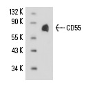
CD55 (143-30) - sc-21769 from Santa Cruz Biotechnology
|
||||||||||
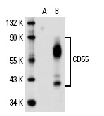
CD55 (H-319) - sc-9156 from Santa Cruz Biotechnology
|
||||||||||
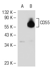
CD55 (67) - sc-53207 from Santa Cruz Biotechnology
|
||||||||||
CD55 (NaM16-4D3) - sc-51733 from Santa Cruz Biotechnology
|
||||||||||
CD55 (H-7) - sc-133220 from Santa Cruz Biotechnology
|
||||||||||
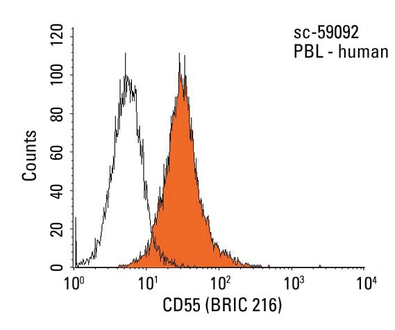
CD55 (BRIC 216) - sc-59092 from Santa Cruz Biotechnology
|
||||||||||
Anti-CD55 antibody - ab96680 from Abcam
|
||||||||||
Anti-CD55 antibody - ab54595 from Abcam
|
||||||||||
Anti-CD55 antibody - ab126221 from Abcam
|
||||||||||
Anti-CD55 antibody - ab136145 from Abcam
|
||||||||||
Anti-CD55 antibody - ab37720 from Abcam
|
||||||||||
Anti-CD55 antibody [143-30] - ab95522 from Abcam
|
||||||||||
Anti-CD55 antibody [143-30] (Biotin) - ab82596 from Abcam
|
||||||||||
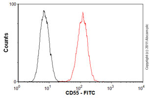
Anti-CD55 antibody [143-30] (FITC) - ab25634 from Abcam
|
||||||||||
Anti-CD55 antibody [143-30] (PE/Cy5®) - ab25410 from Abcam
|
||||||||||
Anti-CD55 antibody [143-30] (Phycoerythrin) - ab25540 from Abcam
|
||||||||||
Anti-CD55 antibody [67] - ab20145 from Abcam
|
||||||||||
Anti-CD55 antibody [BRIC 216] - ab33111 from Abcam
|
||||||||||
Anti-CD55 antibody [EPR6689] - ab133684 from Abcam
|
||||||||||
Anti-CD55 antibody [MEM-118] - ab1422 from Abcam
|
||||||||||
Anti-CD55 antibody [MEM-118] (Alexa Fluor® 488) - ab187775 from Abcam
|
||||||||||
Anti-CD55 antibody [MEM-118] (Biotin) - ab26005 from Abcam
|
||||||||||
Anti-CD55 antibody [MEM-118] (Phycoerythrin) - ab130421 from Abcam
|
||||||||||
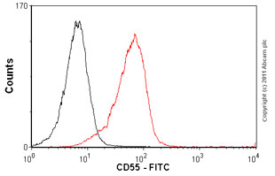
Anti-CD55 antibody [MEM-118], prediluted (FITC) - ab28112 from Abcam
|
||||||||||
Anti-CD55 antibody [MM0175-12L29] - ab89190 from Abcam
|
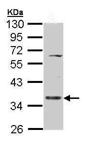
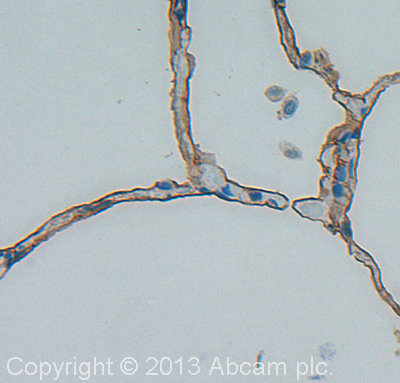
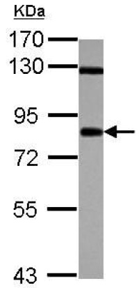
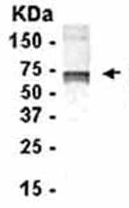
![All lanes : Anti-CD55 antibody [EPR6689] (ab133684) at 1/10000 dilutionLane 1 : A594 cell lysateLane 2 : HeLa cell lysateLysates/proteins at 10 µg per lane.SecondaryHRP labelled goat anti-rabbit at 1/2000 dilution](http://www.bioprodhub.com/system/product_images/ab_products/2/sub_1/25317_CD55-Primary-antibodies-ab133684-1.jpg)
![Human peripheral blood lymphocytes stained with ab1422 (red line). Human whole blood was processed using a modified protocol based on Chow et al, 2005 (PMID: 16080188). In brief, human whole blood was fixed in 4% formaldehyde (methanol-free) for 10 min at 22°C. Red blood cells were then lyzed by the addition of Triton X-100 (final concentration - 0.1%) for 15 min at 37°C. For experimentation, cells were treated with 50% methanol (-20°C) for 15 min at 4°C. Cells were then incubated with the antibody (ab1422, 1μg/1x106 cells) for 30 min at 4°C. The secondary antibody used was DyLight® 488 goat anti-mouse IgM (mu chain) (ab97007) at 1/500 dilution for 30 min at 4°C. Isotype control antibody (black line) was mouse IgM [ICIGM] (ab91545, 1μg/1x106 cells) used under the same conditions. Unlabelled sample (blue line) was also used as a control. Acquisition of >30,000 total events were collected using a 20mW Argon ion laser (488nm) and 525/30 bandpass filter. Gating strategy - peripheral blood](http://www.bioprodhub.com/system/product_images/ab_products/2/sub_1/25321_ab1422-1-ab1422FC.jpg)
